Treatment of Spinal Cord Arteriovenous Malformations
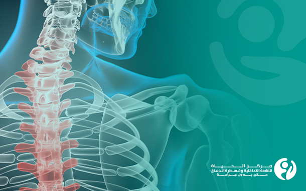
The spinal cord is an extension of the brain within the spinal column. It connects the nerves from the brain to the rest of the body, which allows the brain to control the movements of the limbs and internal organs, such as the bladder or intestines, and receive sensory information like pain or touch from the limbs. We will discuss arteriovenous malformations in the spinal cord and their treatment.
Blood vessels:
Blood vessels are of 3 types: arteries, veins, and capillaries. While arteries carry oxygen-rich blood from the heart to the body, veins return blood to the heart. Capillaries connect the arteries and veins. Any malfunction in the vascular system can cause severe pain, disability, and even death.
Spinal cord arteriovenous malformations diagnosis and treatment:
Vascular malformations in the brain or spinal cord can cause headaches, seizures, or strokes. Diagnosis is usually made by a doctor's examination, and further testing may include imaging tests to provide images of the affected blood vessels and determine what type of vascular malformation the patient has in the central nervous system.
Spinal vascular malformation is a rare condition of an abnormal tangle of blood vessels within or near the spinal cord. It can be congenital, present since birth, or acquired later in life. The malformations can occur spontaneously or due to a tumor. The malformations put increased pressure on the blood vessels and make the patient vulnerable to bleeding or ischemia, depriving the spinal cord of its blood supply, which may lead to nerve injury.
The goal of treating spinal arteriovenous malformations is to block these abnormal connections and restore normal blood flow to the spinal cord. Treatment can reverse or limit the progression of symptoms.
The treatment of spinal arteriovenous malformations (AVMs) at Al Hayat Center for Interventional Radiology and Neurointervention in Iraq focuses on minimally invasive, non-surgical intervention through therapeutic catheterization, which involves making a small incision to allow the injection of medical glue, specialized small coils, or sand-like particles directly into the abnormal vascular connection to close it. The procedure is performed by inserting a small plastic tube into the blood vessels, using high-quality X-ray imaging to guide the tube placement and ensure it is in the correct position before treating the arteriovenous malformation (AVM) or dural arteriovenous fistula (DAVF) with the medical glue, particles, or coils.
What is a spinal cord arteriovenous malformation (AVM)?
Spinal AVM is an abnormal connection between the arteries and veins within or surrounding the spinal cord. Spinal AVMs typically occur in children and younger adults (under 50 years old).
A spinal AVM causes abnormal blood flow within the spinal cord, which can lead to several problems, including internal bleeding and spinal cord stroke, which can result in sudden or gradual loss of limb movement, such as temporary or permanent paralysis, or abnormal sensations like tingling, pins, and needles, or complete loss of sensation in the limbs. There may also be loss of bladder or bowel control. If left untreated, a spinal AVM can progress to severe disability and, though rare, can be fatal.
What does "Dural Arteriovenous Fistula (DAVF) of the Spinal Cord" mean?
A dural arteriovenous fistula (DAVF) of the spinal cord is an abnormal connection between the arteries and veins on the covering (dura) of the spinal cord. DAVF occurs in the elderly, typically after the age of 50.
DAVF causes abnormal blood flow within the spinal cord and can lead to severe spinal disease. There is usually a gradual loss of limb function, such as temporary or permanent paralysis of one or more limbs. There may also be abnormal sensations like tingling, pins and needles, difficulty walking, or loss of limb sensation. There can also be loss of bladder or bowel control, or sexual dysfunction. If left untreated, DAVF can progress to severe disability.
A dural arteriovenous fistula is more insidious and may cause progressive spinal cord dysfunction over many years before diagnosis.
What happens during the treatment of Spinal Vascular Malformations (AVM/DAVF)?
• Spinal vascular malformations are abnormalities of the blood vessels within and around the spinal cord and can be treated under general anesthesia so the patient is asleep and feels nothing.
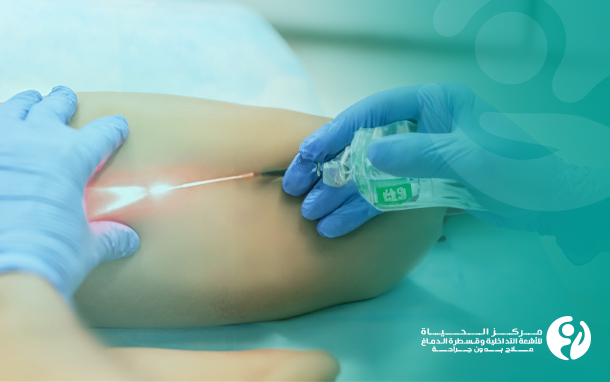
• A small (less than 1 cm) incision is made in the skin of the groin. Through this, a small needle is used to puncture the artery in the groin (femoral artery), and a catheter (a long, thin plastic tube) is inserted into it.
• The interventional radiology and vascular specialist at Al Hayat Center for Interventional Radiology and Neurointervention then carefully navigate this catheter through the blood vessels, using X-ray imaging to guide its placement. The catheter is maneuvered until it is within or near the abnormal blood vessels that will be treated. This is all done without any additional skin incisions beyond the small groin puncture used to insert the catheter.
• Through this catheter, angiography (imaging of the blood vessels) aims to help plan how to block the abnormal blood vessels around the spinal cord to treat the spinal vascular malformation, after capturing and examining images of the blood vessels.
• Radiologists use imaging to determine the required molecules or glue to occlude the blood vessels and how to do it most effectively. The initial planning for this procedure is done during the preparation stage before the day of the procedure, and the final decisions are made after viewing the images from the angiogram.
• Afterward, multiple agents are injected through a catheter into the abnormal connections. These agents work to close off the abnormal connections in the vessels. These agents may be special medical glue, small coils, or sand-like particles, which reduce the severity of the AVM and symptoms and prevent the worsening of the current issues.
• At the end of the procedure, the doctor closes the hole in the femoral artery by internal suturing. The procedure takes 3-6 hours, and the hospital stay is typically 1-2 days.
• Sometimes one treatment session is not enough, so two or three additional sessions may be needed to complete the treatment.
• The treatment outcomes of these conditions are variable and primarily related to the initial degree of impairment and the time interval between the onset and final treatment.
What are the benefits of treating arteriovenous malformations at Al Hayat Center for Interventional Radiology and Neurointervention?
• Reduced risk of future stroke or bleeding.
• Reduction or reversal (fully or partially) of symptoms such as abnormal sensations, limb weakness, urinary incontinence, and loss of bladder/bowel control.
• Reduced need for subsequent surgery.